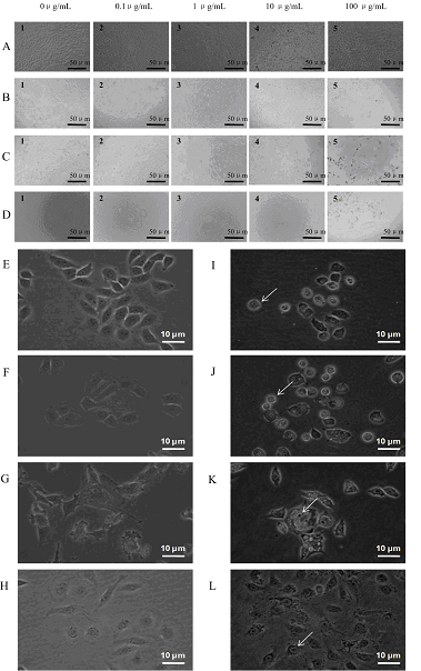
Figure. Morphological characterizations
of liver cell lines.
The four cell
lines were exposed to various doses of TiO2 NPs for 24 hrs and washed with a PBS
solution. The morphology of each cell line was visualized by phase-contrast
microscopy (Nikon, TS 100). (A) SMMC-7721; (B) HL-7702; (C) CBRH-7919; and (D)
BRL-3A. Subgraphs (E)-(H) were the images of SMMC-7721, HL-7702, CBRH-7919, and
BRL-3A cells without NPs, subgraphs (I)-(L) were the images of SMMC-7721,
HL-7702, CBRH-7919, and BRL-3A cells with 100 μg/mL of TiO2, respectively.
Typical cell shrinkage and nuclear condensation were indicated by a white
arrow.
Titanium
dioxide (TiO2) nanoparticles (NPs) are the important nanoscale components of
composites. Although TiO2 NPs and their related nanocomposites have been widely
used in industrial and medical applications, the adverse effects of TiO2
nanomaterials have not been well studied. Here, we investigated the cytotoxicity
of TiO2 NPs in vitro using four liver cell lines: human hepatocellular carcinoma
cell line (SMMC-7721), human liver cell line (HL-7702), rat hepatocarcinoma cell
line (CBRH-7919) and rat liver cell line (BRL-3A). We checked cell viability,
cell morphology, and the levels of reactive oxygen species (ROS) and glutathione
(GSH) after TiO2 exposure at varying concentrations (0.1-100 μg/mL) and
different exposure periods of time (12-48 hrs). Compared to the NP-free control,
all four cell lines exposed to TiO2 NPs showed cytotoxicity in a
dosage-dependent and time-dependent manner, which was associated with the
changes of cell viability and cell morphology, increased intercellular ROS
levels, and decreased intracellular GSH levels. Further, we observed that
carcinomatous liver cells and human liver cells exhibited more tolerance to TiO2
NPs exposure for 24 hrs, compared to normal liver cells and rat liver cells,
respectively. The results indicate that the in vitro cytotoxicity induced by NPs
should be assessed with great caution before the use of nanocomposites and that
there is a need to standardize the cytotoxicity testing procedure of nanoscale
components in composites when using different cell lines.
Title: Cytotoxicity of Titanium Dioxide
Nanoparticles Differs in Four Liver Cells from Human and Rat
Author: BaoYong Sha, Wei Gao, ShuQi
Wang, Feng Xu*, TianJian Lu*
Journal: Composites Part B Engineering
(IF 1.7)
